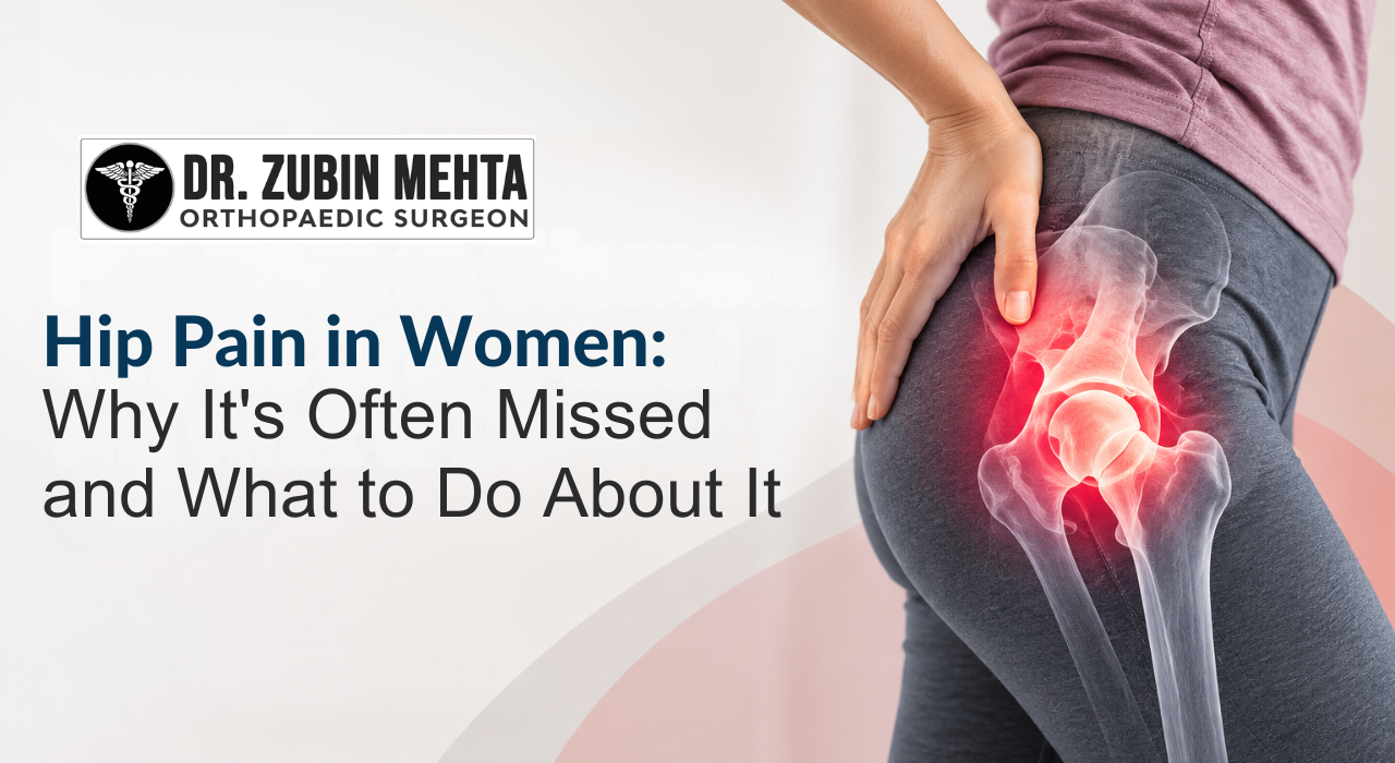
Hip Pain in Women: Why It’s Often Missed and What to Do About It
A pattern I’ve noticed in my OPD over the years: Hip Pain in Women are more likely to arrive late. Not because they ignored the symptoms — but because they were told, at earlier points of contact with the healthcare system, that it was something else.
Lower back problem. Sacroiliac joint. Muscle pull. Gynaecological referral for pelvic pain. Fibromyalgia. The list of misattributions we encounter in the history of female patients with hip pathology is longer than I’d like it to be.
There are reasons for this diagnostic gap — some anatomical, some related to how hip pain presents differently in women, and some, frankly, related to the way pain in female patients is historically under-investigated in medicine. The purpose of this post is to make that gap smaller.
Why Hip Pain Presents Differently in Women
Women have a wider pelvis than men. This anatomical difference — technically a larger Q-angle — affects how forces are transmitted through the hip and knee joints. A wider pelvis means the femoral neck enters the acetabulum at a slightly different angle, which affects how early arthritis, impingement, or labral tears present symptomatically.
The location of pain is also often different. Men with hip osteoarthritis typically describe groin pain clearly and early. Women with the same degree of joint pathology more often describe diffuse thigh pain, lateral hip pain, or referred pain into the buttock. This atypical distribution is frequently misattributed to soft tissue or spinal causes without adequate hip imaging.
Hip Dysplasia: The Condition That Affects Women Far More Than Men
Hip dysplasia — an abnormally shallow hip socket that fails to fully cover the femoral head — affects women at approximately five times the rate of men. It is present from birth and often undiagnosed until adulthood, when it manifests as groin pain, early-onset osteoarthritis, and labral tears.
Mild dysplasia can be managed non-surgically for years, with physiotherapy and activity modification. Significant dysplasia in young women often requires either a periacetabular osteotomy — a bone-repositioning surgery to improve socket coverage — or, if the joint has already developed significant arthritis, a hip replacement. The dysplastic hip replacement is technically more demanding than a standard primary replacement and should be performed by a surgeon experienced with this specific anatomy.
Labral Tears: Common, Commonly Missed
The acetabular labrum is a ring of fibrocartilage around the rim of the hip socket. It deepens the socket, provides a seal that maintains fluid pressure in the joint, and contributes to stability. When the labrum tears — through trauma, repetitive hip motion, or as a consequence of dysplasia or impingement — it causes a distinctive pattern of anterior groin pain, a catching or clicking sensation, and pain with prolonged sitting or hip rotation.
Labral tears do not appear on plain X-ray. They require MRI with arthrogram — an injection of contrast dye into the hip joint before imaging — for reliable diagnosis. If your hip symptoms include anterior groin pain with a clicking or catching sensation, and your plain X-ray is reported as normal, an MRI arthrogram is the investigation you need. Ask specifically for this.
In younger patients without significant arthritis, labral tears can be addressed arthroscopically — through keyhole surgery. In patients with coexisting arthritis, the decision between labral repair and hip replacement depends on the degree of cartilage damage.
Osteoporosis and Hip Fracture Risk
Post-menopausal women face a significantly higher risk of osteoporosis — reduced bone density — which directly increases hip fracture risk. A fall from standing height in a woman with severe osteoporosis can fracture the femoral neck, requiring emergency surgical repair or hip replacement.
The connection to elective hip replacement is this: women approaching the age at which hip replacement may be needed should be assessed for osteoporosis, because bone density affects both surgical timing and implant fixation. Cementless implants — which rely on bone ingrowth for fixation — require adequate bone density. In patients with severe osteoporosis, cemented fixation may be the more appropriate choice.
Every woman over 50 with hip symptoms should have a DEXA scan as part of their pre-operative workup if hip replacement is being considered. This is now part of my standard pre-operative assessment for all female patients over 55.
What to Do If You Suspect Your Hip Has Been Missed
Trust the pattern of your symptoms. Groin pain with weight-bearing that has persisted for more than three months, catching or clicking in the hip with rotation, pain that wakes you at night — these are hip symptoms. They deserve hip imaging: at minimum a plain X-ray of the pelvis and affected hip, and MRI if the X-ray doesn’t explain the severity of your symptoms.
If you’ve been dismissed before, come for a second opinion. Bring all your prior imaging. A thorough clinical examination and the right imaging will usually clarify the diagnosis promptly. Late diagnosis is not inevitable — it is the result of inadequate investigation, and it can be corrected.
CTA: If your hip symptoms have been dismissed or misdiagnosed, come for a proper evaluation. OPD: Tuesday and Thursday, Fortis Hospital Mohali. Call +91 79735 06344.